THORACIC OUTLET ANATOMY

(Hopkins medicine, 2019).
The thoracic outlet consists of the space from the supraclavicular fossa to the axilla. The anatomical margins of the thoracic outlet consist of the interscalene triangle, the costoclavicular space and the subcoracoid space (Chang & Kim, 2021).

(Bassett & Gupta, 2021).
The interscalene triangle is the most medial compartment of the thoracic outlet, the borders are formed by the anterior scalene muscle anteriorly, the middle scalene posteriorly and the medial surface of the first rib inferiorly. The brachial plexus and subclavian artery pass through here (Jones et al., 2019).

(Bassett & Gupta, 2021).
The costoclavicular space is bordered anteriorly by the middle third of the clavicle, posteromedially by the first rib and posterolaterally by the upper border of the scapula. The brachial plexus, sublcavian artery and subclavian vein pass through here (Jones et al., 2019).

(Daniells, 2014).
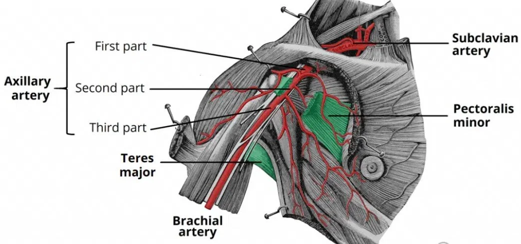
The borders of the subcoracoid space are formed by the pectoralis minor anteriorly, ribs 2-4 posteriorly, and the coracoid process of the scapula. The subclavian artery and vein take on the axillary name as they pass through this space. The cords of the brachial plexus travel throughout this space (Jones et al., 2019).
Neurovascular structureS in tos

(Ortho info, 2023).
The brachial plexus is a complex interplay of nerves formed by the anterior primary rami of the spinal nerve roots of C5-T1. The fibers of the nerve roots combine to form the brachial plexus trunks found in the posterior triangle of the neck, between the anterior and middle scalene muscles. The C5 and C6 nerve roots form the superior trunk, C7 continues as the middle trunk and C8 and T1 form the inferior trunk.
The trunks of the brachial plexus split into anterior and posterior divisions, which form the medial, lateral, and posterior cords. This subdivision of the brachial plexus contains five major terminal branches, musculocutaneous, axillary, radial, median, and ulnar nerves which provide motor and sensory innervation to the upper extremity (Polcaro et al., 2023).

(Kenhub, 2019).
The subclavian vein is a major venous channel that is located beneath the clavicle and drains deoxygenated blood from the upper limb, head, and neck and returns it to the heart. The subclavian vein lies anterior and inferior to the subclavian artery and travels medially to the sternoclavicular joint. From here it amalgamates with the internal jugular vein to form the brachiocephalic vein (Capobianco et al., 2023).
(Kenhub, 2019).
The subclavian artery is a major artery that supplies blood to the upper extremities, head and neck. The right side originates from the brachiocephalic trunk which is a branch of the aortic arch. The left side originates directly from the aortic arch after the left common carotid artery. The subclavian artery progresses laterally beneath the clavicle, over the first rib and to the axilla. The artery is split to three parts dependent on its correlation with the anterior scalene muscle. The first part is proximal, second part is posterior and the third part is distal to the anterior scalene muscle. The distal aspect continues as the axillary artery once it crosses the lateral border of the first rib (Rahimi & Geiger, 2023).